Cells built complex nanoscale ‘machines’ from basic biomolecular building blocks to perform vital biological functions. These cellular ‘machines’ are at the heart of key processes in mechanobiology, such as cell migration, cell adhesion, and mechanotransduction. Our overarching research goal is to gain a comprehensive insight into the nanoscale structure-function relationship that governs the assembly, organization, dynamics, and functions of these cellular machines. Our approach is highly interdisciplinary, combining advanced imaging technologies with rigorous molecular and cell biology methods.
Super-resolution microscopy
We have pioneered the use of superresolution microscopy to elucidate nanoscale architecture of cellular structures (Kanchanawong et al., Nature, 2010), and have long-standing involvement in the development of ultra-high resolution 3D imaging techniques, iPALM (Shtengel et al., PNAS 2009). Our focus is in advancing the capability of super-resolution microscopy, using several platforms including our own iPALM system and surface-generated structured illumination techniques.
Bioimage informatics
Super-resolution and advanced microscopy techniques generate beautiful, complex, and exquisitely detailed images of cells in large quantity. These images contain vast amount of information but it is still very challenging to quantitatively, rigorously, and comprehensively analyse such datasets. To fully tap the potential of these 21st century imaging techniques, this analysis bottleneck must be tackled. We have several ongoing projects where we seek to leverage computer vision and machine learning approaches to unlock information contained in super-resolution microscopy images (for example: Zhang et al., MBoC 2017).
Focal adhesions
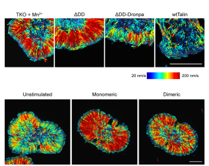
Focal adhesions are major cell adhesion structures that mediate cell-extracellular matrix (ECM) adhesions. Focal adhesions play essential roles in mechanotransduction, rigidity sensing, and cell migration. Our focus is in understanding the molecular architecture of focal adhesions (Kanchanawong et al., Nature, 2010) and how they are animated during cellular functions. Our recent work established the roles of the protein Talin as the determinant of focal adhesions architecture (Liu et al., PNAS 2015). Ongoing projects seek to combine nanoscale imaging with molecular engineering approaches to understand the operational principles that control focal adhesions structure and functions.
Cell-cell junctions
In tissues, coherent organization of cells depends on cadherin-mediated cell-cell junctions. We have recently elucidated for the nanoscale architecture of cadherin-based cell adhesions, using superresolution microscopy (Bertocchi et al., Nature Cell Biology, 2017; Wu et al., Developmental Cell, 2015). In our ongoing projects we seek to understand comprehensively the transformation and linkages between nanoscale structures and functions during the formation and maturation of epithelial tissues